


The Wonders of Brain Imaging by Kaja Posnik
- cellfiemagazine

- May 29, 2021
- 4 min read
Welcome back #STEMinists. How are you all? We hope that you enjoyed seeing our part of the Cambridge Science Festival last month and we cannot wait to share with you our new ventures going forward. Today, our monthly blog writer Kaja has researched the fascinating world of brain imaging. Thank you for your hard work, Kaja.
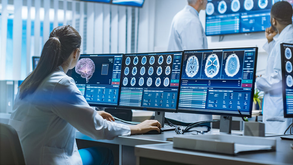
Have you ever wondered what is needed to diagnose diseases that are often invisible at first sight? Or how they can be treated if there is still so much unknown about them? Even though the field is still developing and a lot more research has to be done to be confident in understanding and approaching them, what can be used to learn more about invisible illnesses such as mental disorders are brain imaging techniques. Brain scans, especially Magnetic Resonance Imaging (MRI) and Functional Magnetic Resonance Imaging (fMRI) have recently become more frequently used to help mental disorder patients receive an appropriate diagnosis and hope for a better future.
Magnetic Resonance Imaging is a brain scanning technique, which shows the anatomy and structure of the brain in a three dimension in a safer way than using traditional X-Rays. MRI is non-invasive and relies on a strong magnetic field created by a magnet embedded in the machine. The field acts on the protons in the human body, changing their spin and aligning them with the direction of the magnetic field. When the magnet is turned off and the magnetic field stops influencing the protons, they return back to their original position. The energy released by their going back to normal and the time it takes to do this is measured and based on that researchers know what the molecules are and what type of tissue is being investigated.
A version of MRI is fMRI - Functional Magnetic Resonance Imaging, which in turn is able to show the activity and functioning of the brain, not just its structure. It is a Blood Oxygen Level Dependent technique (BOLD), which measures brain activity based on the amount of oxygen that is delivered to an area of the brain. When a patient is performing a cognitive task in the machine, the activated area requires more blood flow and therefore oxygen, which is carried in oxyhaemoglobin. The active part of the brain attracts more oxygenated blood, which is also more detectable by the machine and gives a stronger signal than deoxygenated blood, therefore showing which area is responsible for certain functioning. fMRI has a high spatial resolution, resulting in detailed images, however as it has a poor temporal resolution (often gives the result of the scanning with a slight delay - after the brain activity had happened) it may not be the best method to use in the detection of all illnesses and brain damages.
Getting back to the idea of using these brain imaging techniques to diagnose mental illnesses, advances have been made in the field with the help of neuromarkers. Scientists have identified the patterns that various mental health disorders present when the brain is scanned and they can compare the ones obtained from patients to the distinguished neuromarkers. This enables researchers to identify activity particular to e.g. schizophrenia or depression and give the patient a diagnosis based on the similarity between the detected pattern and the one of the neuromarker. Although this is still not an ideal method of diagnosis, as the more illnesses the machine is programmed to compare, the more difficult it gets for it to differentiate between them, it is a good step in the direction of improvements in the field of mental illnesses.
Unlike patient interviews, therapy and other more subjective methods of mental illness diagnosis, which, although could be successful, may not provide clear scientific evidence of such disorders, brain scans are a more objective way of showing distorted brain structure or faulty functioning. Neuroimaging, for instance, can differentiate between the symptoms caused by a tumour in the frontal lobe and a mental disorder such as depression which can affect e.g. the amygdala and hippocampus. Brain scans could even help predict whether a child will suffer from mental health problems in the future, as proposed by researchers led by professor Susan Whitfield-Gabrieli. Such advances could eventually help prevent the disorders instead of treating them and biomarkers, again, could play a very big role in making that possible.
All in all, despite the fact that the use of neuroimaging to detect mental health disorders and help find treatment for them is still a developing field, a lot of hope is put into it. Although brain scans may not always be able to differentiate between the illnesses nor be appropriate for diagnosing patients who suffer from multiple disorders at the same time or may only be successful at detecting mental disorders at their later stage, after they had done some damage, they can still open many doors into more successful and objective diagnosis. By basing it on facts and images of brain structure and functioning, the patients could be provided with more information, which could hopefully enable them to receive better suited and more appropriate treatment during the course of the disorder.
Sources:


Comments